Reinforced Cardiac Patch
Working with cardiac surgeons to make a regenerative cardiac patch
The Clinical Problem
Cardiac patches are used to repair defects in the heart wall, including ventricular septal defects. Current materials, such as PTFE, Dacron, or bovine pericardium, are strong and implantable, but biologically inert. They do not integrate, degrade, or contribute to heart function, meaning they stabilise the defect without restoring tissue. While regenerative approaches aim to address this, they are not yet clinically viable and lack the mechanical robustness required for implantation.
The Approach
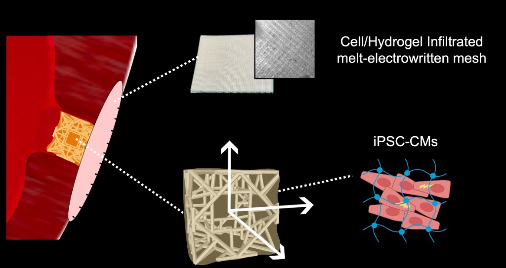
The core challenge was combining mechanical robustness with a viable cellular environment in a single component. A cardiac patch must withstand pressure, hold sutures, prevent bleeding, and support living cells — requirements that are difficult to achieve together. Instead of relying on a single material, we combined complementary 3D-printed components into a single implant. A 3D printed lattice provides structural support, while a mesh enables suturing and prevents bleeding. This structure can then be infiltrated with a cell-laden matrix containing cardiomyocytes, making the patch both implantable and regenerative.
What We Built
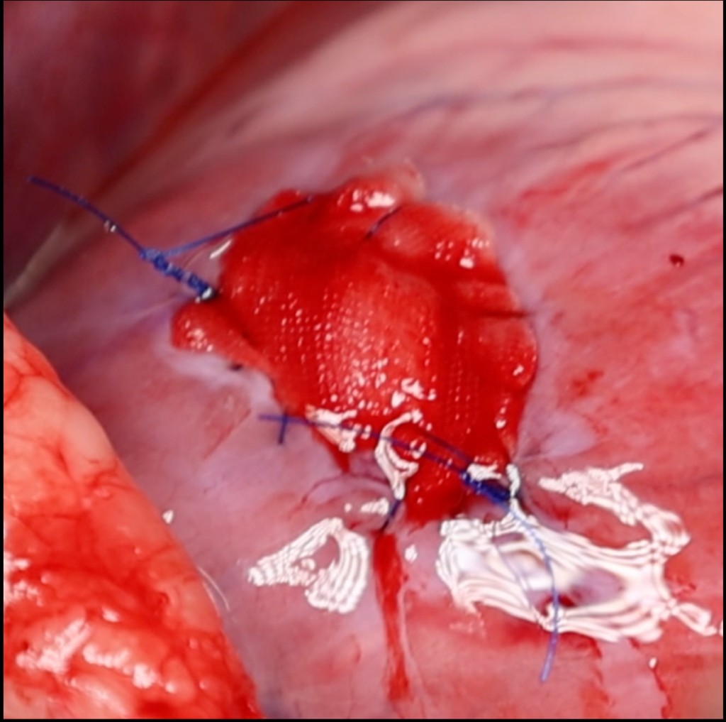
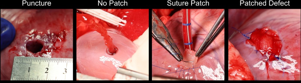
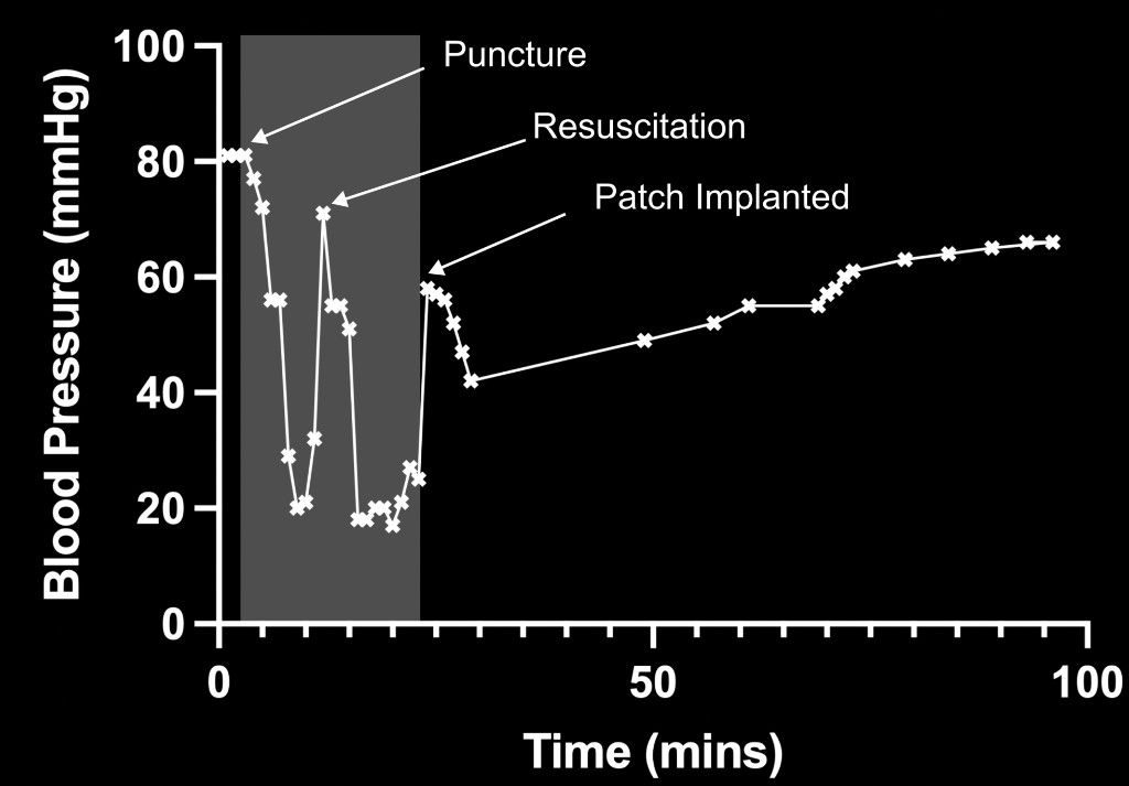
Our multi-material cardiac patch combines a 3D-printed lattice, a melt-electrowritten mesh, and a cell-laden hydrogel. We demonstrated that the patch has material properties (stiffness) that match the heart and can support 3D cell growth. Furthermore, we showed that the patch can be implanted via suturing, prevents bleeding, and withstands heart contraction. We implanted the patch in a porcine model; it was sutured into a ventricular defect and withstood pressures of ~100 mmHg. The patch sealed the defect without sustained bleeding within minutes, and remained stable on a beating heart for over 90 minutes.
My Role
I led the project from concept to publication, working across engineering, biology, and surgical translation. I defined the technical approach with clinical collaborators, coordinated the design and fabrication strategy, and developed the experimental pipeline. I also designed the in vivo study and supported the surgical implantation to ensure the system could be evaluated under realistic conditions. I wrote and led the publication process.
Outcomes
This work demonstrates that advanced manufacturing techniques — volumetric printing and melt electrowriting — can be used to engineer a cardiac patch that is both implantable and biologically functional. Furthermore, it demonstrates the effectiveness of microstructured materials in reinforcing soft, fragile, cell-laden materials. In the future, these fabrication techniques could be used in other regenerative medicine applications.
Related publications
- First authorVolumetric 3D Printing and Melt-Electrowriting to Fabricate Implantable Reinforced Cardiac Tissue PatchesAdvanced Materials · 2025
- First authorPreprintVolumetric 3D Printing and Melt-Electrowriting to Fabricate Implantable Reinforced Cardiac Tissue PatchesbioRxiv · 2025
Related media
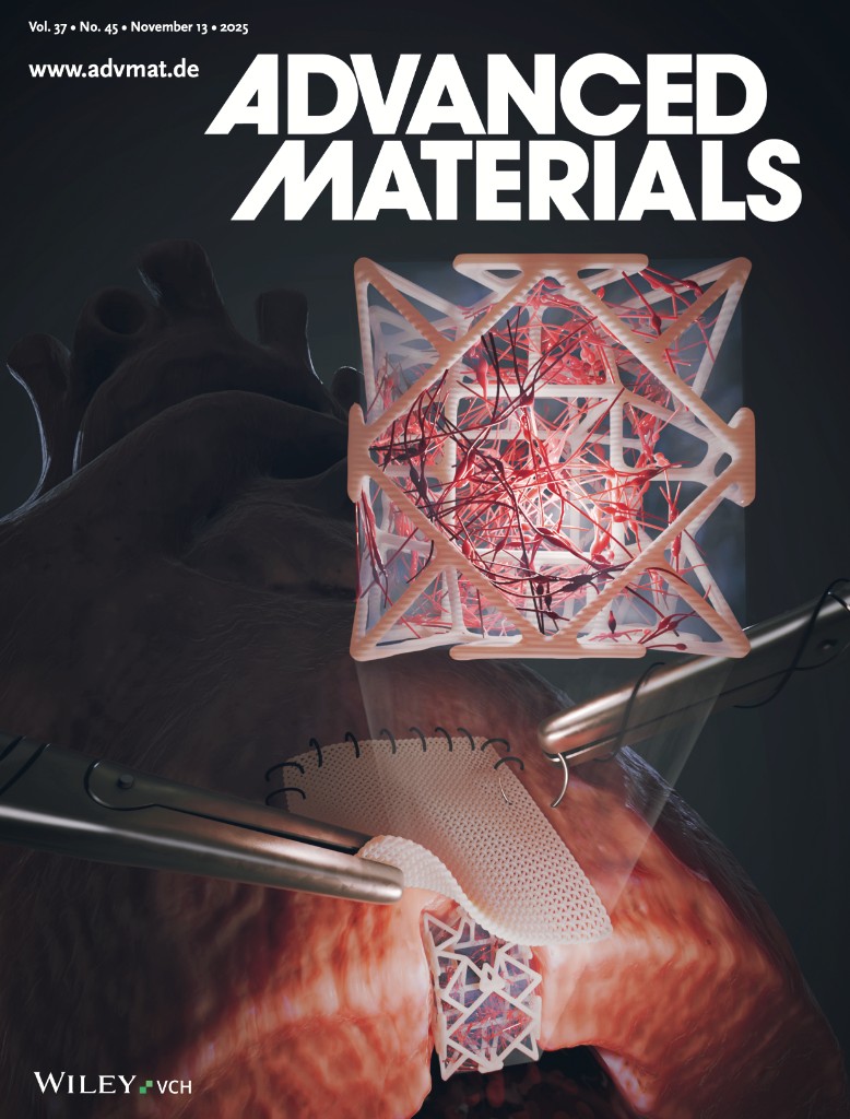
Advanced Materials Cover Art
My work was selected to be the cover piece in Adv. Mater. 45/2025
Reinforced Cardiac Tissue
ETH press release video related to the reinforced cardiac patch.